
Scapular dyskinesia: alteration of the scapula movement
Scapular dyskinesia is the loss of normal synchrony in the scapular-thoracic joint complex. This causes an alteration of the position and normal movements of the scapula. This alteration can be the cause of anatomical imbalances in structures adjacent to the scapula or in the scapula itself.
How to diagnose Scapular Dyskinesia?
According to the symptoms manifested by the patient during the physical evaluation, the application of functional tests starts in order to identify if the problem really has scapular origin or comes from the shoulder joint complex, so the evaluation must be very exhaustive. Three important aspects should also be taken into account:
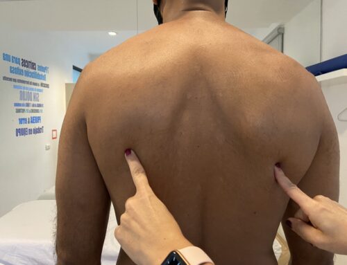
- Evaluation through clinical observation on the patient’s body posture to know if that is Scapular Dyskinesia.
- The effect of manual correction in relation to the symptoms.
- Evaluation of the scapula itself and adjacent anatomical structures; through functional tests, to define which are responsible for the observed dyskinesia.
Classification of Scapular Dyskinesia
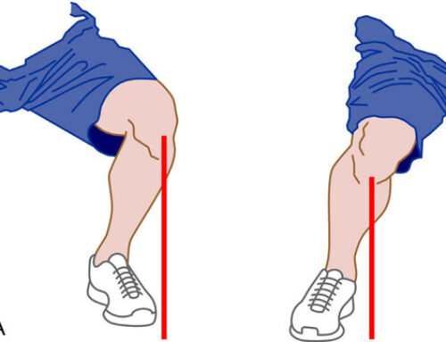
Scapular dyskinesia can be divided into types, depending on the result after the patient is asked to raise both arms. There are 3 types:
Causes
- Soft tissue stiffness, often found in the pectoralis minor muscle and levator scapulae, could lead to inadequate scapular tilt.
- Possible neuromuscular fatigue and scapulothoracic incoordination. A modification of the motor strategy resulting in compensatory muscle activity will likely lead to muscle overload.
- The movement of the shoulder girdle involves the sternoclavicular, acromioclavicular and glenohumeral joints, usually moving all simultaneously.
- Functional defects of any of the joints impairs the movements of the shoulder girdle.
- Postural alterations that promote this pathology, as in a resting position with excessive thoracic kyphosis and increased cervical lordosis.
- The contracture of the posterior capsule or retraction of the posterior stabilizing complex of the glenohumeral joint which can generate an alteration in the kinematics of the glenohumeral joint.
When talking about scapular dyskinesia, it should be clear that in most cases the cervical region and the shoulder may be involved, so it enters into a controversy because the real cause of pain can be a glenohumeral or cervical origin.
Taking into account what has been described above, it is important to do a global evaluation of the upper body, that means that the entire upper limb and the rest of the anatomical structures of the scapula, shoulder and neck should be included.
For this reason, it is extremely important to carry out the analysis and rehabilitation with specialists.
Scapular dyskinesia is the loss of normal synchrony in the scapular-thoracic joint complex. This causes an alteration of the position and normal movements of the scapula. This alteration can be the cause of anatomical imbalances in structures adjacent to the scapula or in the scapula itself.
How to diagnose Scapular Dyskinesia?
According to the symptoms manifested by the patient during the physical evaluation, the application of functional tests starts in order to identify if the problem really has scapular origin or comes from the shoulder joint complex, so the evaluation must be very exhaustive. Three important aspects should also be taken into account:
- Evaluation through clinical observation on the patient’s body posture to know if that is Scapular Dyskinesia.
- The effect of manual correction in relation to the symptoms.
- Evaluation of the scapula itself and adjacent anatomical structures; through functional tests, to define which are responsible for the observed dyskinesia.
Classification of Scapular Dyskinesia
Scapular dyskinesia can be divided into types, depending on the result after the patient is asked to raise both arms. There are 3 types:
Causes
- Soft tissue stiffness, often found in the pectoralis minor muscle and levator scapulae, could lead to inadequate scapular tilt.
- Possible neuromuscular fatigue and scapulothoracic incoordination. A modification of the motor strategy resulting in compensatory muscle activity will likely lead to muscle overload.
- The movement of the shoulder girdle involves the sternoclavicular, acromioclavicular and glenohumeral joints, usually moving all simultaneously.
- Functional defects of any of the joints impairs the movements of the shoulder girdle.
- Postural alterations that promote this pathology, as in a resting position with excessive thoracic kyphosis and increased cervical lordosis.
- The contracture of the posterior capsule or retraction of the posterior stabilizing complex of the glenohumeral joint which can generate an alteration in the kinematics of the glenohumeral joint.
When talking about scapular dyskinesia, it should be clear that in most cases the cervical region and the shoulder may be involved, so it enters into a controversy because the real cause of pain can be a glenohumeral or cervical origin.
Taking into account what has been described above, it is important to do a global evaluation of the upper body, that means that the entire upper limb and the rest of the anatomical structures of the scapula, shoulder and neck should be included.
For this reason, it is extremely important to carry out the analysis and rehabilitation with specialists.